What Are the 2026 Top Orthopaedic Medical Devices?

Orthopaedic medical devices will enter 2026 under strong clinical and demographic pressure. The World Health Organization reports that approximately 1.71 billion people live with musculoskeletal conditions worldwide. Osteoarthritis, fractures, back pain, and sports injuries continue to influence hospital demand, rehabilitation needs, and implant innovation.

Market forecasts support this direction. Fortune Business Insights estimates that the global orthopaedic devices market reached about USD 42.5 billion in 2024 and may expand substantially by 2032. Grand View Research also identifies rising joint-replacement procedures, ageing populations, and improved surgical technologies as major growth drivers. These figures are useful, but forecast methods differ. Market size should not be treated as clinical proof.

This 2026 overview examines the leading device categories through evidence, usability, safety, and patient outcomes. It considers robotic-assisted systems, hip and knee implants, spinal devices, trauma fixation products, sports-medicine tools, and orthobiologics. A surgeon may judge a device by handling and visibility in a crowded operating room. A patient may care more about walking downstairs without pain. Both perspectives matter.

The strongest products are not always the newest. They should demonstrate reliable design, appropriate regulatory clearance, clear labeling, and meaningful follow-up data. The U.S. FDA, peer-reviewed studies, registry findings, and manufacturer clinical evidence provide important reference points. Still, evidence gaps remain, especially for emerging digital and robotic platforms. That uncertainty deserves attention.

This guide therefore avoids a simple popularity contest. It asks a harder question: which orthopaedic medical devices offer the clearest value for real clinical practice in 2026?

What Are the 2026 Top Orthopaedic Medical Devices?

Global Burden: 1.71 Billion People Need Musculoskeletal Care (WHO)

What Are the 2026 Top Orthopaedic Medical Devices?

Global Burden: 1.71 Billion People Need Musculoskeletal Care (WHO)

The World Health Organization reports that 1.71 billion people live with musculoskeletal conditions worldwide. This burden includes osteoarthritis, fractures, back pain, and sports-related injuries. These conditions affect mobility, employment, independence, and daily comfort. The number is not abstract. It can mean a painful knee on stairs or a worker unable to lift safely.

In 2026, leading orthopaedic devices will likely focus on joint replacement systems, trauma fixation implants, spinal devices, and surgical navigation tools. Advanced implants increasingly use porous structures and patient-specific designs. These features may support bone integration and improve surgical accuracy. The Global Burden of Disease Study 2019 identified low back pain as the leading cause of disability globally. That evidence strengthens demand for safer spinal solutions and earlier rehabilitation.

Digital rehabilitation devices will also gain attention. Motion sensors can measure walking symmetry, joint angles, and recovery progress outside hospitals. The OECD’s Health at a Glance reports continuing pressure on healthcare capacity and ageing populations. Remote monitoring may help clinicians manage follow-up care more efficiently. Still, better technology does not guarantee better outcomes. Not every patient needs a smarter implant. Cost, training, repair access, and unequal healthcare systems remain serious concerns. The field still has blind spots. A device can be technically impressive yet unsuitable for a patient’s lifestyle, anatomy, or local clinical resources.

Sources: World Health Organization, Musculoskeletal Health; Global Burden of Disease Study 2019, The Lancet; OECD, Health at a Glance.

Joint-Replacement Devices: Knee Demand May Reach 3.48 Million by 2030

What Are the 2026 Top Orthopaedic Medical Devices?

Knee replacement devices are expected to remain a major orthopaedic category through 2026. Market projections suggest annual demand could reach 3.48 million procedures by 2030. That figure reflects ageing populations, longer life expectancy, and rising mobility expectations. It is still a projection, not a guaranteed outcome. Local access, surgical capacity, and reimbursement rules may change the pace.

Modern knee systems commonly include femoral and tibial components, a bearing insert, and a patellar component. Advanced designs aim to restore alignment, reduce wear, and support natural movement.

Computer-assisted navigation can help surgeons plan bone cuts and check implant positioning during surgery. Robotic assistance may improve consistency, but it does not replace clinical judgment. Patient anatomy still matters most.

Small details matter. A surgeon may review standing X-rays, walking patterns, ligament stability, and pain location before choosing a device. Materials must tolerate repeated loading inside a warm, active body. Long-term evidence should include revision rates, function, and patient-reported outcomes.

Faster recovery sounds attractive. However, recovery varies widely between patients. Some early studies also have limited follow-up, leaving important questions unanswered. Device selection should therefore remain individualized, transparent, and based on trained medical evaluation rather than market excitement.

Spinal Implants: Devices Addressing the Rising Burden of Back Pain

What Are the 2026 Top Orthopaedic Medical Devices?

Spinal implants are gaining attention as back pain affects work, sleep, and daily movement. In 2026, leading device categories include interbody cages, pedicle screw systems, artificial discs, and motion-preserving implants. These devices support stability, restore disc height, or reduce pressure on irritated nerves. Some use porous surfaces that encourage bone growth. Others aim to preserve controlled spinal movement. The “best” implant depends on anatomy, diagnosis, bone quality, and surgical goals. No device fits every patient.

Modern planning increasingly combines imaging, navigation, and minimally invasive techniques. Surgeons can assess alignment before surgery and guide instruments through smaller openings. This may reduce tissue damage, but it does not remove surgical risks. Infection, nerve injury, nonunion, implant loosening, and adjacent-segment problems remain possible. Long-term evidence also varies between implant types. That matters. A promising design still needs careful clinical follow-up, transparent data, and approval for its intended use.

Tips: Ask what problem the implant is designed to solve. Request evidence for similar patients, not only laboratory results. Discuss bone density, recovery time, revision risks, and realistic activity limits. A second opinion can clarify difficult choices. Keep imaging records and attend follow-up visits, even when symptoms improve. Patients sometimes focus on the device and overlook rehabilitation. That is a mistake worth reconsidering.

Trauma Fixation Systems: Plates, Screws, Nails, and External Frames

What Are the 2026 Top Orthopaedic Medical Devices?

Trauma fixation systems remain central to orthopaedic care in 2026. Plates, screws, intramedullary nails, and external frames support different fracture patterns. Each device must match the bone, injury, and patient’s healing potential. A low-profile plate can stabilize an ankle fracture while limiting soft-tissue irritation. Locking screws may help in weak bone, but they do not replace accurate reduction. Small errors still matter.

Intramedullary nails provide strong load-sharing support for many long-bone fractures. Their alignment depends on careful entry-point preparation and reliable imaging. Surgeons often value shorter procedures, although speed should never outweigh positioning accuracy. External frames remain valuable when swelling, contamination, or severe soft-tissue damage limits internal fixation. They can stabilize a limb quickly. Pin-site care remains essential, and patient adherence is sometimes underestimated.

Material selection also deserves attention. Titanium alloys may reduce imaging distortion, while stainless steel offers familiar strength and handling characteristics. Biocompatibility, fatigue resistance, instrument design, and sterilization workflows influence real-world performance. Clinical evidence should guide device selection more than marketing language. No system is perfect. Some newer designs appear promising, yet long-term comparative data may still be limited. Teams should review fracture classification, vascular status, bone quality, and postoperative rehabilitation before choosing a construct. A technically advanced implant can fail when follow-up is inconsistent or the fixation plan ignores the patient’s daily demands.

What Are the 2026 Top Orthopaedic Medical Devices? - Trauma Fixation Systems: Plates, Screws, Nails, and External Frames
Device Category Device Type Common Implant Materials Primary Fixation Principle Typical Trauma Applications Representative Design Features Key Clinical Advantages Important Considerations
Plates Compression Plate Titanium alloy or stainless steel Applies compression across a fracture through eccentric screw placement or plate contouring. Simple transverse or short-oblique fractures of long bones, including selected diaphyseal injuries. Round or oval holes; can be contoured to match the bone surface; available in narrow and broad profiles. Provides direct fracture compression and strong resistance to bending. Requires adequate soft-tissue coverage and may reduce periosteal blood supply when extensively exposed.
Plates Locking Compression Plate Titanium alloy or stainless steel Combines conventional compression with fixed-angle screw fixation. Comminuted fractures, osteoporotic bone, periarticular fractures, and metaphyseal injuries. Threaded plate holes accept locking screws; combination holes may permit both locking and non-locking fixation. Creates a stable fixed-angle construct and reduces dependence on plate-to-bone friction. Correct reduction remains essential; excessive stiffness or poor working-length selection can affect callus formation.
Plates Periarticular Anatomical Plate Titanium alloy or stainless steel Supports the joint-adjacent fragment while maintaining fixed-angle or conventional fixation. Distal femur, proximal tibia, distal tibia, proximal humerus, distal radius, and other periarticular fractures. Precontoured geometry, multiple multidirectional screw options, and low-profile designs. Improves control of small articular or metaphyseal fragments and helps restore alignment. Accurate plate positioning is important to avoid joint penetration, tendon irritation, or malalignment.
Plates Clavicle and Small-Bone Plate Titanium alloy or stainless steel Neutralization, compression, or bridge fixation according to fracture pattern. Displaced clavicle fractures, forearm fractures, hand fractures, and foot fractures. Low-profile shapes, limited-contact undersurfaces, and holes designed for smaller-diameter screws. Allows precise fixation in anatomically small or subcutaneous regions. Prominence-related discomfort and soft-tissue irritation may occur, particularly in areas with limited coverage.
Screws Cortical Screw Titanium alloy or stainless steel Threads engage dense cortical bone to provide axial compression or plate fixation. Diaphyseal fixation, plate attachment, and interfragmentary lag fixation when appropriate. Fine, closely spaced threads and a relatively small thread depth. Effective purchase in dense cortical bone and broad compatibility with plate systems. Appropriate length and bicortical purchase must be confirmed; over-tightening can damage bone or strip threads.
Screws Cancellous Screw Titanium alloy or stainless steel Engages trabecular bone with deeper, wider-spaced threads and may provide interfragmentary compression. Metaphyseal and epiphyseal fractures, malleolar fractures, and selected articular fragments. Large thread depth, partial- or full-thread options, and compatibility with washers where needed. Designed for purchase in softer cancellous bone and can compress selected fracture fragments. Thread purchase may be limited in severely osteoporotic bone; joint penetration must be avoided.
Screws Cannulated Screw Titanium alloy or stainless steel Guided over a guidewire for accurate placement, with compression generated by screw design and tightening. Femoral neck, malleolar, radial head, pelvic, and other fractures requiring controlled screw trajectories. Central cannulation, partially or fully threaded configurations, and optional washers. Facilitates minimally invasive, image-guided placement and accurate trajectory control. Guidewire position must be monitored; cannulated screws may have lower core strength than equivalent solid screws.
Intramedullary Nails Locked Intramedullary Nail Titanium alloy or stainless steel Load-sharing fixation within the medullary canal, supplemented by proximal and distal locking screws. Femoral and tibial shaft fractures, and selected humeral shaft fractures. Various diameters and lengths, interlocking holes, end caps, and options for static or dynamic locking. Preserves fracture-site biology compared with extensive plating and supports early mobilization. Entry-point accuracy, canal preparation, rotation, alignment, and locking-screw placement are critical.
Intramedullary Nails Cephalomedullary Nail Titanium alloy or stainless steel Intramedullary load sharing combined with a large proximal element that supports the femoral head and neck. Intertrochanteric and subtrochanteric proximal femur fractures. Short or long nail options, a lag screw or helical blade, and distal locking capability. Provides a mechanically favorable construct for many unstable proximal femur fractures. Correct reduction and head-neck element position are necessary to reduce cut-out, malrotation, and implant-related complications.
Intramedullary Nails Flexible Intramedullary Nail Typically titanium alloy or stainless steel Elastic bending forces provide relative stability and maintain alignment in selected patients. Selected pediatric long-bone fractures, especially femoral and forearm fractures. Small-diameter flexible nails inserted through metaphyseal entry points, commonly used as paired implants. Minimally invasive placement with limited disruption of the fracture hematoma. Less suitable for highly unstable patterns or heavier patients; nail balance and end positioning are important.
External Frames Unilateral External Fixator Stainless steel or titanium half-pins with aluminum or carbon-fiber connecting components Load is transferred through percutaneous pins or wires to an external frame. Temporary stabilization, open fractures, severe soft-tissue injury, damage-control orthopaedics, and selected definitive treatments. Unilateral bars, clamps, adjustable pin positions, and modular frame configurations. Rapid application with access to wounds and soft tissues; useful when internal fixation is unsafe. Pin-site infection, frame stability, alignment maintenance, and patient tolerance require ongoing management.
External Frames Circular Ring Fixator Stainless steel or titanium wires and half-pins with circular metal or composite rings Uses tensioned wires and/or half-pins connected to rings to provide multiplanar stability. Complex open fractures, periarticular injuries, bone loss, infection-related reconstruction, and deformity correction. Multiple rings, tensioned wires, adjustable struts, and computer-assisted or manual frame adjustments. Allows multiplanar control, gradual deformity correction, and access to compromised soft tissues. Application is technically demanding; requires prolonged follow-up, pin-site care, and patient compliance.
External Frames Hybrid External Fixator Metal rings or partial rings combined with tensioned wires and half-pins Combines circular fixation near the joint with half-pin fixation in the diaphysis. Proximal tibia, distal tibia, distal femur, and other fractures with limited periarticular bone stock. Partial rings, tensioned wires, threaded half-pins, and modular connecting rods. Provides strong periarticular fixation while preserving access to the joint and soft tissues. Wire or pin trajectory must avoid neurovascular structures, tendons, and the joint capsule.
Note: The specifications and applications shown are representative clinical characteristics of trauma fixation technologies. Exact dimensions, compatibility, sterilization requirements, and indications vary by device system and applicable regulatory instructions.

Digital Orthopaedics: Robotics, Navigation, Sensors, and 3D Printing

What Are the 2026 Top Orthopaedic Medical Devices?

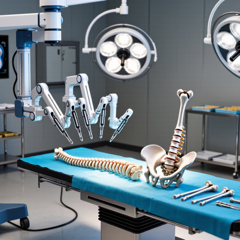
Digital orthopaedics is moving from isolated tools toward connected surgical workflows. The International Federation of Robotics reported about 6,100 medical robots sold globally in 2023, a 36% increase from the previous year. In orthopaedic theatres, robotic systems can support bone preparation, alignment checks, and repeatable instrument positioning. Navigation adds real-time anatomical guidance, while optical or electromagnetic trackers monitor small movements. Accuracy matters. However, a precise system cannot correct poor imaging or weak surgical planning.

Smart instruments are also becoming more practical. Force sensors can detect resistance during drilling, cutting, or implant preparation. Pressure sensors may help assess balance around a joint. These measurements could reduce guesswork, but sensor readings still require clinical interpretation. A 2024 industry analysis from MarketsandMarkets projected strong growth for surgical navigation and robotic-assisted surgery through the decade. Market forecasts are useful, though they do not prove better patient outcomes.

Three-dimensional printing supports patient-specific guides, anatomical models, and selected implants. The Wohlers Report 2024 valued the global additive manufacturing industry at about 20.0 billion dollars in 2023. Hospitals can print a knee model, inspect difficult geometry, and rehearse the surgical approach before incision. That is tangible. Yet printed planning tools may increase workflow time, validation duties, and training demands. I have seen digital precision treated as clinical certainty. That remains a mistake. Data quality, surgeon experience, cybersecurity, and long-term outcome tracking still deserve equal attention.

The chart compares representative positioning or dimensional-accuracy benchmarks commonly reported for major digital orthopaedic technologies. Lower values indicate smaller deviation. These values are technology-level reference ranges rather than company or brand measurements.